Phased Arrays and the Beating Heart: How Acoustic Interference Is Redefining Cardiac Imaging
The human heart completes roughly 100,000 contraction cycles every day, each one a coordinated mechanical event involving electrical propagation, fluid dynamics, and tissue deformation occurring across multiple spatial scales simultaneously. Capturing that event with sufficient fidelity to detect subtle pathology has been the central engineering challenge of cardiac diagnostics for decades. Increasingly, the answer lies not in brute-force improvements to transducer sensitivity but in the deliberate and sophisticated manipulation of acoustic interference—a strategy that transforms the physics of wave superposition into clinical intelligence.
The Transducer Array as an Interference Engine
Conventional single-element ultrasound operates on a straightforward principle: emit a pulse, receive the echo, and reconstruct depth from time-of-flight. The spatial resolution of such a system is fundamentally constrained by the wavelength of the emitted sound and the aperture of the transducer. For cardiac applications—where millimeter-scale features can carry life-or-death diagnostic significance—these constraints are not merely inconvenient; they are clinically limiting.
Phased-array transducers resolve this limitation by replacing the single element with a dense grid of individual emitters, each of which can be driven with an independently controlled phase delay. When these elements fire in a precisely sequenced pattern, their emitted wavefronts interfere constructively along a steerable focal axis and destructively in all other directions. The result is a focused acoustic beam that can be electronically swept across the cardiac field of view in milliseconds—no mechanical moving parts, no interruption between frames.
The interference geometry here is exact and deliberate. If the inter-element spacing is $d$ and the desired beam steering angle is $\theta$, the required phase delay between adjacent elements is $\Delta\phi = (2\pi d \sin\theta)/\lambda$, where $\lambda$ is the acoustic wavelength in tissue. By varying $\Delta\phi$ in real time, the array can steer the constructive interference maximum to any desired angle within the transducer's field of view, effectively painting a two-dimensional image in a single cardiac cycle.
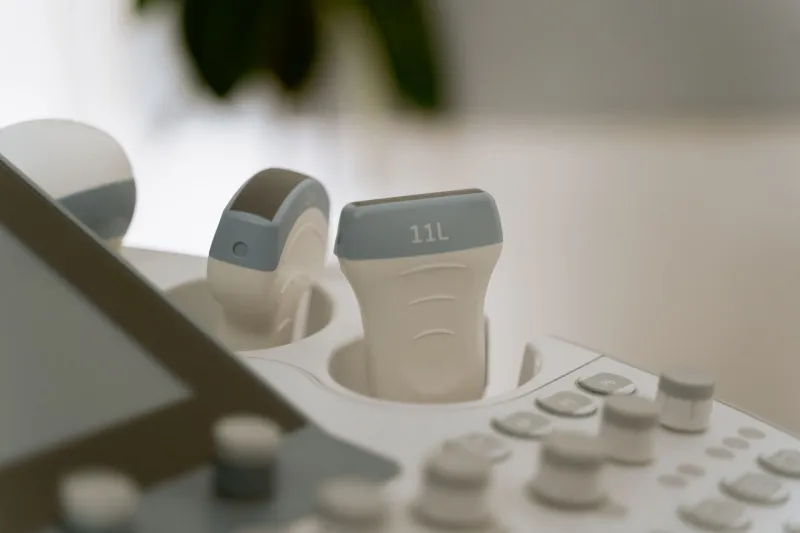
Modern cardiac transducers used in clinical echocardiography—such as those deployed in matrix-array probes by manufacturers including Philips, GE HealthCare, and Siemens Healthineers—extend this principle into three dimensions, with element counts ranging from 2,000 to over 9,000 individual piezoelectric elements arranged in a two-dimensional grid. The interference patterns they generate are correspondingly complex, capable of forming volumetric focal zones that update at frame rates sufficient to capture rapid events such as valve leaflet motion during systole.
Coherent Compounding: Averaging Interference for Clarity
One of the most consequential recent advances in cardiac ultrasound is spatial coherent compounding, a technique that borrows directly from synthetic aperture radar and sonar. Rather than acquiring a single focused beam per image line, compounding fires multiple unfocused or weakly focused plane waves at different steering angles and coherently combines the resulting echo datasets.
The word "coherently" is critical. Coherent summation preserves phase information, which means that echoes from true reflectors—tissue interfaces, valve structures, myocardial walls—add constructively across the combined frames, while acoustic noise and clutter, which lack consistent phase relationships, tend to cancel through destructive interference. The signal-to-noise ratio of the compounded image therefore scales favorably with the number of compounding angles, yielding images with substantially improved contrast resolution compared to conventional focused beamforming.
For cardiologists, improved contrast resolution translates directly into diagnostic capability. Endocardial border definition—the visibility of the inner surface of the heart wall—is a persistent challenge in echocardiography, particularly in patients who are obese or have hyperinflated lungs, conditions that are prevalent in the American patient population. Coherent compounding has been shown in multiple clinical studies to improve endocardial visibility in such patients, reducing the rate of non-diagnostic examinations and decreasing reliance on contrast agents.
Detecting the Subtle: Arrhythmias and Structural Defects
Perhaps the most clinically compelling application of interference-based ultrasound advances is in the detection of pathology that conventional echocardiography routinely misses. Two categories are particularly illustrative.
First, subtle structural defects such as small ventricular septal defects, atrial septal aneurysms, and early hypertrophic cardiomyopathy changes involve tissue abnormalities on the order of one to three millimeters. At standard clinical ultrasound frequencies of two to five megahertz, the diffraction-limited resolution of a conventional focused beam is insufficient to reliably characterize such features. High-frequency matrix arrays operating at ten to fifteen megahertz, combined with coherent compounding, can achieve lateral resolutions approaching 0.3 millimeters in near-field cardiac windows—sufficient to visualize septal morphology with a degree of detail previously requiring transesophageal approaches.
Second, functional arrhythmia detection using ultrasound has historically been indirect—clinicians infer rhythm disturbances from electrocardiographic data and then use echocardiography to assess mechanical consequences. Emerging high-frame-rate ultrasound systems, capable of acquiring volumetric data at rates exceeding 1,000 frames per second using unfocused plane wave transmissions and parallel receive beamforming, now permit direct visualization of electromechanical wave propagation across the myocardium. These so-called electromechanical wave imaging (EWI) techniques map the interference patterns created by the interaction of propagating activation fronts with regional myocardial stiffness variations, effectively rendering the electrical activation sequence visible as a mechanical wave. Research groups at Columbia University and the University of California San Francisco have demonstrated the technique's capacity to localize ectopic pacemaker foci and characterize re-entrant pathways in patients with ventricular tachycardia—information that previously required invasive electrophysiology studies.
Clinical Translation and the Road Ahead
The translation of these interference-based techniques into routine clinical practice in the United States is proceeding along multiple fronts. The American Society of Echocardiography has begun incorporating three-dimensional volumetric assessment into its recommended protocols for structural heart disease evaluation, a shift that implicitly endorses phased-array interference beamforming as a standard of care. Major academic medical centers, including the Cleveland Clinic, Mayo Clinic, and Massachusetts General Hospital, have deployed next-generation matrix-array systems for research and clinical use.
Computational demands remain a meaningful barrier. Coherent compounding of volumetric data from thousands of array elements in real time requires processing architectures that push the limits of current GPU-based systems. Several research groups are exploring field-programmable gate array implementations and neural network-accelerated beamforming to reduce latency, with the goal of making high-frame-rate three-dimensional compounding available on portable and point-of-care devices—a development that could extend advanced cardiac imaging to emergency departments and rural clinical settings where specialist echocardiographers are unavailable.
The heart, in its ceaseless mechanical rhythm, is itself a kind of wave generator—pressure pulses propagating through a fluid-filled elastic chamber. It is fitting, then, that the most powerful tools for understanding its pathology are built on the same fundamental physics: waves interfering, canceling, and reinforcing, yielding information from the superposition of signals that no single pulse alone could provide.