Productive Noise: How Researchers Are Weaponizing Interference Patterns to Sharpen Cancer Imaging
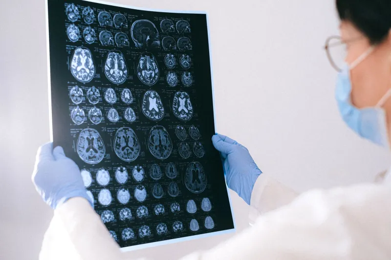
The conventional wisdom in diagnostic imaging is straightforward: noise is the enemy. Signal-to-noise ratio is the metric by which imaging systems are evaluated, improved, and marketed. Radiologists are trained to interpret images in spite of noise, and engineers are rewarded for suppressing it. The cleaner the signal, the better the diagnosis — or so the logic has run for decades.
A quieter, more technically precise conversation is now taking place in research hospitals and signal processing laboratories across the country, and it challenges that assumption at a fundamental level. Under specific conditions, interference — including what registers as noise in conventional imaging frameworks — can be structured, exploited, and directed to reveal pathological features that clean signals routinely miss. The counterintuitive result is that some tumors are more visible, not less, when the imaging system introduces controlled wave interference into the detection process.
Rethinking Signal Purity
To understand why, it helps to revisit what noise in medical imaging actually represents at the wave level. In ultrasound systems, the received signal is a superposition of echoes returning from tissue boundaries at varying depths and acoustic impedances. What manifests as speckle — the granular texture visible in ultrasound images — is not random contamination. It is the product of coherent interference among multiple backscattered wavefronts arriving at the transducer simultaneously.
Speckle has historically been treated as a degradation artifact, and considerable engineering effort has been devoted to reducing it through spatial compounding, frequency averaging, and adaptive filtering. The assumption underlying these approaches is that speckle obscures real anatomical structure. In many contexts, that assumption is correct. But research emerging from institutions including the University of Michigan and Stanford's medical imaging groups has identified conditions in which speckle patterns carry diagnostic information that smoothing algorithms discard along with the noise.
Small lesions — particularly those in early-stage malignancy, when their acoustic properties differ only subtly from surrounding tissue — can produce interference signatures within the speckle field that are detectable through statistical analysis of the interference pattern itself, even when the lesion is too small or too acoustically similar to its environment to produce a distinguishable echo on its own.
Constructive Interference as a Sensitivity Mechanism
The more deliberately engineered application of this principle involves transmit-side manipulation — shaping the outgoing wave field to create constructive interference at target tissue locations. Coherent compounding techniques, originally developed to reduce speckle through incoherent averaging, have been adapted by several research groups to do the opposite: to deliberately induce constructive interference at suspected lesion sites by transmitting plane waves at multiple angles and combining the resulting echoes with phase-aligned summation.
When the phase relationships are tuned correctly, the technique concentrates acoustic energy at the target location with an efficiency that focused single-beam imaging cannot match. The constructive interference effectively amplifies the echo from tissues that would otherwise fall below the system's detection threshold. In phantom studies simulating early-stage breast lesions, phase-coherent compounding has demonstrated sensitivity improvements of 15 to 30 percent over conventional B-mode imaging — a margin that, in clinical terms, corresponds to the difference between detecting a 4-millimeter mass and missing it entirely.
The mechanism is, at its core, the same constructive interference principle that governs phased-array radar and acoustic beamforming. The innovation lies in applying it to the receive-side reconstruction of tissue echoes rather than simply to transmit-side beam steering.
MRI and the Noise Floor as a Diagnostic Layer
In magnetic resonance imaging, the interference dynamics operate differently but yield analogous insights. MRI signal acquisition is a phase-sensitive process: the received signal encodes spatial information through deliberate phase modulation, and the reconstruction algorithms that produce diagnostic images are, fundamentally, interference-based computations — inverse Fourier transforms that extract spatial information from the superposition of phase-encoded signals.
Thermal noise in MRI, arising from random electromagnetic fluctuations in both the patient's tissue and the receiver coil electronics, has traditionally been minimized through hardware improvements and signal averaging. But researchers working with ultra-high-field MRI systems — 7 Tesla and above, now available at select academic medical centers in the US — have observed that at these field strengths, the interference between the transmitted radiofrequency field and tissue-induced standing waves creates spatial variations in flip angle and signal intensity that were initially treated as artifacts.
Subsequent analysis revealed that these interference-induced variations carry information about tissue dielectric properties — properties that differ measurably between healthy and malignant tissue. Tumors, particularly those with elevated water content and altered ion concentrations characteristic of rapidly dividing cells, produce distinctive dielectric signatures that manifest as predictable perturbations in the RF interference pattern. Groups at the University of Minnesota and NYU Langone have developed reconstruction algorithms that extract these dielectric maps from the interference data, adding a layer of tissue characterization that conventional magnitude-only MRI images do not provide.
Stochastic Resonance: When Random Noise Helps Weak Signals
Perhaps the most theoretically striking application of interference-informed imaging involves a phenomenon borrowed from nonlinear systems theory: stochastic resonance. In stochastic resonance, the addition of a precisely calibrated level of random noise to a system carrying a subthreshold signal can enhance the detection of that signal — not despite the noise, but because of it.
The mechanism depends on interference between the noise floor and the signal at the nonlinear detection threshold of the system. When noise amplitude is tuned to the right level, it intermittently pushes weak signals above the detection threshold in a temporally structured way that preserves the signal's information content. Add too little noise and the signal remains buried. Add too much and it is overwhelmed. At the optimal noise level, the interference between signal and noise produces a detection probability that exceeds what either could achieve independently.
Researchers at MIT's Research Laboratory of Electronics have demonstrated stochastic resonance enhancement in ultrasound contrast agent detection, a technique relevant to perfusion imaging in tumor vasculature characterization. By introducing controlled noise into the receive signal chain at levels calibrated to the expected contrast agent echo amplitude, they achieved statistically significant improvements in microbubble detection sensitivity — a result that required abandoning the assumption that noise reduction is always the correct engineering objective.
Clinical Translation and the Road Ahead
Translating interference-enhanced imaging from research settings to routine clinical practice involves regulatory, workflow, and interpretive challenges that the technical results alone cannot resolve. The FDA's clearance pathway for imaging algorithms requires clinical validation data that most interference-exploitation techniques have not yet accumulated at scale. Radiologists trained on conventional image appearance will require recalibration when presented with interference-derived tissue maps that look nothing like familiar anatomical renderings.
Nevertheless, the trajectory is clear. Several interference-informed ultrasound algorithms are already embedded in commercial systems under the marketing language of "advanced compounding" or "tissue characterization," their wave-physics underpinnings rarely explained to end users. As ultra-high-field MRI becomes more accessible and photoacoustic imaging — which is inherently an interference-based modality — moves toward clinical deployment, the productive exploitation of wave interference in diagnostic medicine will become less a research curiosity and more a standard engineering assumption.
The signal, it turns out, was hiding in the noise all along.